Immunoassays

Flow Cytometry

Accurate & Precise Analyses

Flow cytometry, commonly referred to as FACS analysis, is an effective method when used to identify and measure cellular biomarkers in complex subpopulations. Scorpius utilizes the Beckman Coulter CytoFLEX S flow cytometry platforms as part of our premier immunoassay facilities. Flow cytometry allows for accurate and precise analyses of cell surface and intracellular molecules to better characterize and define cell types in a mixed or purified sample.

Measuring Cells Individually

A key advantage of flow cytometry is its ability to analyze and measure cells individually. This is particularly beneficial when studying heterogeneous populations of cells, as it can be used to rapidly analyze subpopulations in just a few minutes, producing very detailed data on count, properties, and classification on a cell-by-cell basis. The Beckman Coulter CytoFLEX S is especially valuable for high-throughput, high-accuracy cell counting, as well as for applications involving biomarker detection. Scorpius' scientists are skilled in a broad variety of flow cytometry applications in support of non-GLP, GLP and cGMP product release testing.
cta_autoMACS@2x

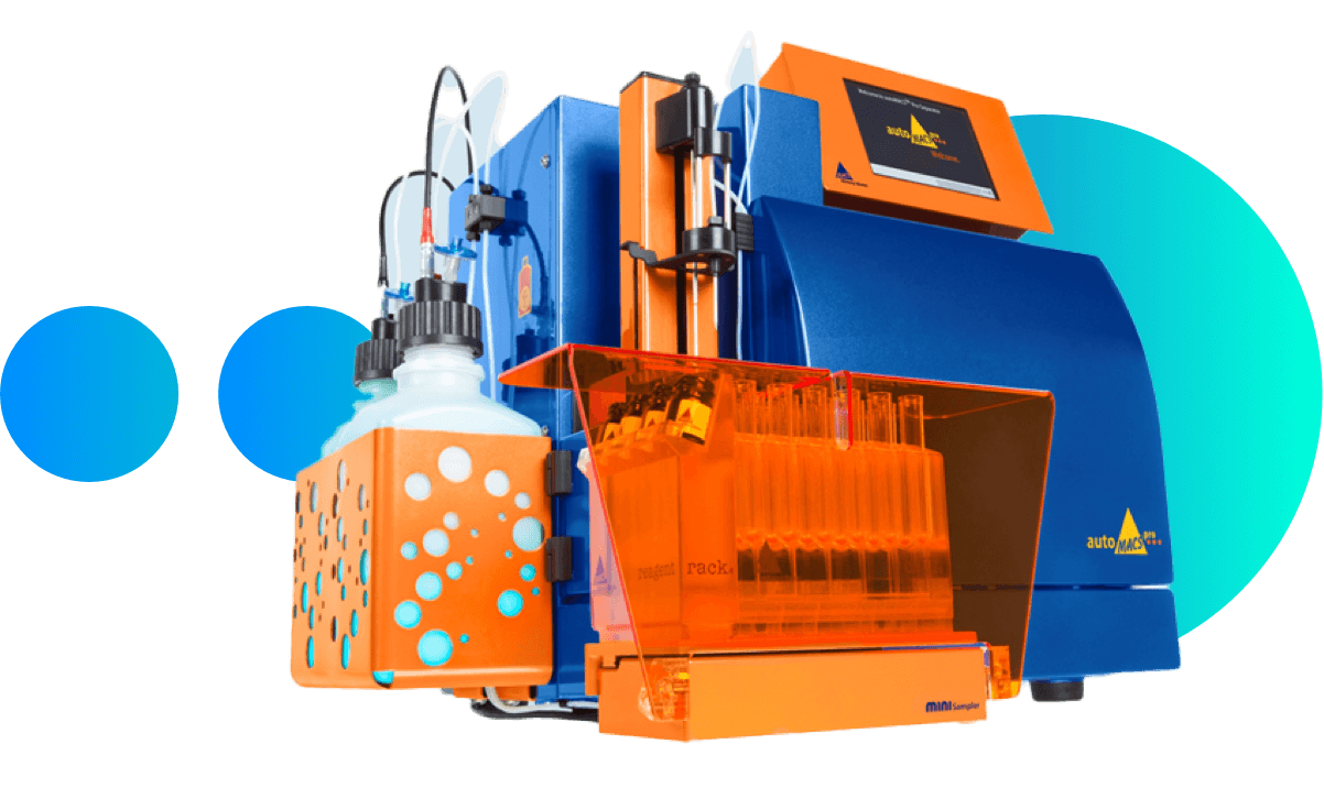
Scorpius' autoMACS® Pro Separator

The autoMACS® Pro Separator enables fast and gentle isolation of virtually any cell type, ideal for downstream applications like flow cytometry.

Our Technologies
Scorpius has qualified the following panels to assess proliferative activity or expression levels of immune checkpoint-related molecules and T cell population:

Memory panel:

CD3, CD4, CD8, CD197, CD45RA, CD27, CD57, PD-1, CD56

Exhaustion markers:
Regulatory cells:
1. Class II Tetramer Assay

Measuring the ability of cells to respond to synthetic antigens and/or whole proteins in response to tetanus toxoids. This application facilitates the study of antigen specific CD4 T cells.

2. Antigen-Specific T Cell Response

T cell panel optimized for the expression of effector cytokines, including TNF gamma, IFN gamma, granzyme B, and perforin.

3. Apoptosis Assay

This assay is utilized to detect and quantify cellular events leading to apoptosis in an individual population of live cells. Early stages of apoptosis can be detected by the activation of caspase enzymes, which are labeled with a bright fluorescent signal. This can then be analyzed on the CytoFLEX Flow Cytometer to determine the percent of live, necrotic, and apoptotic cells.